This meeting is designed to talk about the current challenges in developing and using super-resolution microscopy. With short talks and lots of time for discussion, the workshop will discuss recent advances in super-resolution imaging from new developments in imaging to analysis of super-resolution data. We particularly want to encourage early career researchers to attend and contribute to the meeting. Please contact the organisers if you would like more information on how to contribute.
University of Leeds
University of Leeds
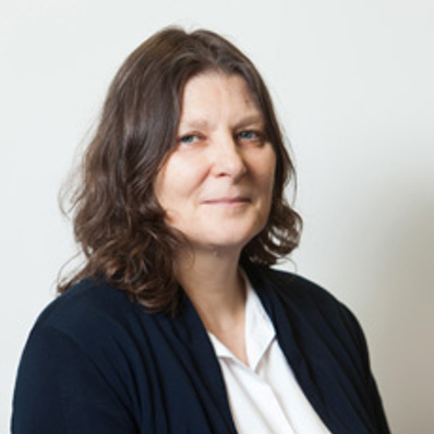
Aleks obtained a PhD in Mechanical Engineering at Imperial College London, where he applied novel fluorescence microscopy techniques to study confined fluids. He then moved to the Chemistry department in Cambridge, where he worked on new single-molecule and light-sheet microscopy tools for investigating the behaviour and organisation of membrane proteins in T cells. In 2020, Aleks starts his own lab as a University Academic Fellow at the Bragg Centre for Materials Research in Leeds. Here, his lab will focus on the application of high-speed fluorescence imaging to push beyond the temporal limits of single-molecule and super-resolution fluorescence microscopy.
University of Leeds
University of Leeds
Michelle is Professor of Cell Biology in the Faculty of Biological Sciences. She obtained a BA in Physiology of Organisms at the University of York, and a PhD in Physiology at University College London. She moved to King's College London, and started to use a specialised form of light microscopy (birefringence) to investigate muscle crossbridge orientation. She then worked at UCSF, San Francisco for a year, where she used fluorescence polarisation to investigate muscle crossbridges. She moved back to the UK, to the University of York, to work on insect flight muscle. In 1990 she was awarded a Royal Society University Research fellowship, based at King's College London, and began working on the cell and molecular biology of muscle development, and started to use live cell imaging to investigate muscle cell behaviour in cultured cells, and confocal microscopy to investigate their cytoskeleton. She collaborated with Graham Dunn to use Digitally Recorded Interference Microscopy with Automatic Phase Shifting (DRIMAPS) to investigate cell crawling behaviour. She moved to Leeds in 1997 as a Lecturer, and has continued to use a wide range of both light and electron microscopy approaches to investigate the molecular motors and the cytoskeleton.
Alex is a Postdoctoral Research Fellow at the University of York. He specializes in studying biomolecular condensates, and their capillary and single-molecule physics, using fluorescence coupled with optical tweezers. These condensates have diverse functions, underpinning antimicrobial resistance, managing biological memory, and even contributing to carbon fixation.
His background is in physical chemistry and optics, but during his PhD at Durham University, he developed into a multidisciplinary light microscopist and image analyst. He has subsequently designed and delivered projects ranging from a clinical trial on human fertility, to postdoctoral work in cardiac imaging, and a JSPS Fellowship focused on molecular cell signalling in cancer. He constantly seeks new models, organisms, and condensates to investigate and finds ways to measure their behavior on the smallest scales possible with light.
He is a leader for the representation of ECR scientists at York and informs strategy for improved Research Culture. He also advocates for Open Research standards in light microscopy through the QUAREP-LiMi network.
Understanding the epigenetics of flowering with SlimVar single molecule tracking Friday @ 11:10 AM
University of Cambridge, UK
Research in TheLeeLab centers on developing new biophysical methods to answer fundamental biological questions, primarily through single-molecule fluorescence spectroscopy and multidimensional super-resolution imaging. Professor Lee completed his DPhil in Physical Chemistry with Dr. Mark Osborne, before postdoctoral work with both Prof Sir David Klenerman (FRS MedSci) and Prof W.E Moerner (Nobel Prize Chemistry 2014). He manages a research team of talented researchers who profoundly believe molecules should be looked at one at a time. Professor Lee is the 2017 recipient of the Marlow Prize in Physical Chemistry from the Royal Society of Chemistry.
Multidimensional Super-resolution Imaging: Wasting Light to Learn New Things Friday @ 10:45 AM
University of Strathclyde, UK
University of StrathclydeSuper-resolution imaging using the Mesolens Friday @ 1:10 PM
Aisha Haneea Syeda is a molecular biologist and biophysicist with extensive research and teaching experience specialising in DNA metabolism and genome stability. She employs cutting-edge single-molecule techniques to explore the fundamental metabolic processes that occur on DNA with a particular focus on the impact of DNA-protein complexes on genome stability. By investigating the interplay of these molecular processes, Aisha aims to uncover mechanisms that contribute to genome stability. The insights gained from her work have the potential to advance cancer therapies and develop innovative antibiotics to address the growing problem of antibiotic resistance.
In vivo single molecule microscopy reveals that replisomes stack and disassemble at barriers to DNA replication Friday @ 1:35 PM
King's College London
Dr Susan Cox works at the Randall Centre for Cell and Molecular Biophysics, developing fluorescence microscopy techniques and applying them to discover new cell biology at the nanoscale. In 2011 she was awarded a Royal Society University Research Fellowship, which she used to develop a substantial research program based around localisation microscopy, and methods to extract more information from super-resolution image data. SC is best known as the developer of Bayesian analysis of blinking and bleaching (3B), a method for analysing extremely dense localisation microscopy image series. Its importance has been recognised with the award of the Royal Microscopical Society light microscopy medal and the Society of Experimental Biology Presidents Medal. More recently, she has explored the limits of localisation in terms of speed and accuracy. She mathematically described the role of the size of the point spread function size in limiting information transmission speed and developed a machine learning based approach to remove poor fits from the super resolution image. Since it is obviously more desirable to avoid poor fits in the first place, she developed Haar Wavelet Kernel analysis (HAWK), an approach to localisation microscopy data analysis which avoids artifacts and ensures the results reflect the underlying structure of the sample.
Deep learning for data synthesis in fluorescence microscopy Friday @ 3:15 PM
Sandeep holds an MSci in Theoretical Physics and Applied Mathematics, followed by a PhD in Mathematical Biology from the University of Birmingham. Currently, she is a Research Fellow in Immunology and Immunotherapy at the University of Birmingham, where her work focuses on applying deep learning techniques to analyse the spatial organisation of proteins across various conditions, utilising Single-Molecule Localization Microscopy (SMLM), a super-resolution imaging method. Her research interests include mathematical modelling of biological systems through differential equations, parameter estimation, and machine learning, with a particular emphasis on deep learning applications in biological data analysis.
An omics-type approach to compare spatial organisation of proteins within cells using deep learning Friday @ 3:40 PM
Tagging proteins for live-cell super-resolution imaging Friday @ 1:50 PM
CAIRN
CAIRN Research are an independent, employee owned microscopy research-orientated company specialising in solutions for the life sciences for nearly 40 years. From their origins as a provider of turnkey optical systems for fast intracellular ion measurements, we have maintained close links with our academic customer base. This has enabled them to develop a broad range of products and solutions to meet their expanding needs. CAIRNS's strength lies in thier understanding of both the requirements of the microscopy research community and of the advances in technology from which they can benefit.
CAIRN Research are instrument designers, manufacturers and vendors of both Illumination and Detection products. Their illumination products encompass a wide range of light sources, adapters and specialist laser systems; typically used in conjunction with a third-party, or Cairn-built, microscope or macrosope. On the detection side, the primary focus is on multi-channel imaging using one or more third-party cameras. These products are largely based around optical techniques, most importantly, fluorescence, optogenetics, photolysis and transmitted light imaging.
Although fully independent they are closely allied with Chroma Technology where a large number of our illumination and detection products make use of their high performance interference filters.
CAIRN Friday @ 4:30 PM
University of York
Sarah is a PhD student in the University of York Physics of Life group supervised by Steven Quinn and Mark Leake. She is studying the effects of molecular crowding and other environmental changes on biomolecules using single-molecule FRET microscopy techniques. Specifically work so far has focused on DNA hairpins and their opening and closing dynamics and the SARS-CoV-2 pseudoknot which acts as the stimulatory sequence for frameshifting in the virus.
Shining light on nucleic acid dynamics with smFRET Friday @ 4:00 PM
Photon Lines
Photon Lines Ltd was incorporated in May 2003, and since then has become a leading UK and Ireland scientific distributor with an impressive portfolio of products.
Our customers include the physics, chemistry and biology departments of world-leading universities, the Medical Research Council, CRUK, and government research facilities including the Diamond Light Source, STFC and AWE. Many blue-chip manufacturing facilities are using our camera based products, including high speed cameras and long-term recording systems for trouble shooting on production lines and laser beam profiler/power meters for 100% testing of active components used in high volume manufacturing of consumer electronics products
With sister offices in France and Spain, Photon Lines Ltd is able to call upon a pool of expertise in the field of photonics to the benefit of customers and suppliers alike.
Photon Lines Ltd is ISO Accredited to comply with the Quality standard ISO9001:2015.
Photon Lines UK is a small but highly qualified team specialising in photonics applications in Life and Physical Sciences.
Photon Lines Friday @ 4:15 PM
Sahil is a PhD candidate in the Department of Physics at IIT Delhi and a Commonwealth Split-Site Scholar at the University of Glasgow. His research is dedicated to advancing reconstruction and illumination techniques for structured illumination microscopy.
Absorption modulation based non-linear SIM Friday @ 11:40 AM
Jack Shepherd is a postdoctoral researcher at the University of York, where he uses super resolution microscopy, single molecule force transduction tools, and molecular dynamics to probe systems ranging from single molecule DNA structure and dynamics to the algal pyrenoid to molecular crowding in yeast. He is principally interested in creating novel equipment, software, and methods including the magneto-optical tweezing fluorescence microscope COMBI-Tweez, and Python based single molecule tracking and diffusion/stoichiometry estimation software package PySTACHIO.
Using Super-Resolution Techniques to Estimate the Size, Position, and Mobility of DNA Plectonemes Friday @ 11:50 AM
Oliver Umney is a final year PhD student at the UKRI Centre for Doctoral Training in Artificial Intelligence for Medical Diagnosis and Care at the University of Leeds.
In his project, Oliver has been looking at whether the organisation of the ligand epiregulin, imaged via SMLM, could predict response to anti-EGFR treatment in metastatic colorectal cancer patients.
To do this he has been developing machine-learning pipelines for analysis of SMLM data, which he hope may be useful to others in the community.
Segmentation and classification of SMLM data using deep-learning Friday @ 2:05 PM
Newcastle University
Dr Kevin Whitley is a Lecturer in the Centre for Bacterial Cell Biology at Newcastle University. Research in his new lab primarily focuses on understanding the molecular mechanism of bacterial cell wall biosynthesis using single-molecule and super-resolution methods. His PhD (University of Illinois Urbana-Champaign) involved using optical tweezers and single-molecule fluorescence to investigate the elasticity of nucleic acids in vitro, but then as a postdoc he decided to start looking at single molecules in live (bacterial) cells. Kevin's postdoc work (Newcastle University and TU Delft) focused on investigating the molecular mechanism of bacterial cell division using a combination of nanofabrication, microfluidics, and single-molecule microscopy. With his own young group, he is investigating the biosynthesis of cell wall biosynthesis and cell division in the bacterium Corynebacterium glutamicum, a model system for the large and understudied phylum Actinobacteria.
Divided we stand: Watching bacterial cell division one molecule at a time using vertical cells Friday @ 11:25 AM
The RMS Ambassador scheme Friday @ 3:55 PM