- You are here:
- Homepage
- Library
- Journal of Microscopy
- Editors & Editorial Board
Editors

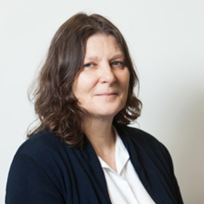
General Editor, Michelle Peckham
School of Molecular and Cellular Biology, University of Leeds, UK,
Expertise in confocal, deconvolution and super-resolution imaging, transmission and scanning electron microscopy and cryo-electron microscopy.

General Editor, Michelle Peckham
School of Molecular and Cellular Biology, University of Leeds, UK,
Expertise in confocal, deconvolution and super-resolution imaging, transmission and scanning electron microscopy and cryo-electron microscopy.
Michelle Peckham obtained her degree in Biology from the University of York in 1981 and her PhD from University College London in 1984. She then spent 2 years at KCL Biophysics working on muscle birefringence, followed by a year at the University of San Francisco, working on fluorescence polarisation microscopy, and then 2 years at the University of York, working on insect flight muscle contraction. She became a Royal Society University Research Fellow in the department of Biophysics at KCL in 1990, when she set up her own group working on muscle and cytoskeleton, and as part of that work, she began using confocal microscopy. She moved to the University of Leeds in 1997, and became Professor of Cell Biology in 2010. She uses a number of different imaging techniques in her work, from confocal, deconvolution and super-resolution imaging, to transmission and scanning electron microscopy, as well as cryo-electron microscopy. She was president of the Royal Microscopical Society from 2016 – 2019.

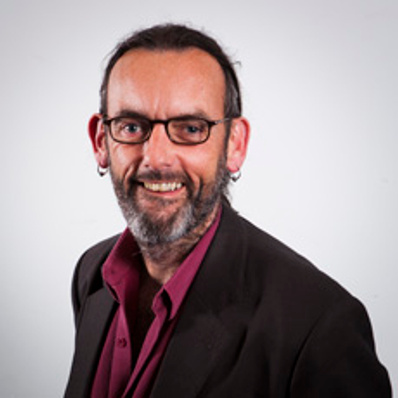
Deputy Editor, Peter Nellist
Department of Materials, University of Oxford, UK,
Expertise in transmission electron microscopy and scanning transmission electron microscopy imaging, diffraction and spectroscopy techniques and applications.

Deputy Editor, Peter Nellist
Department of Materials, University of Oxford, UK,
Expertise in transmission electron microscopy and scanning transmission electron microscopy imaging, diffraction and spectroscopy techniques and applications.
Pete Nellist is a Professor in the Department of Materials, and a Tutorial Fellow at Corpus Christi College, University of Oxford. He leads a research group that focuses on the applications and development of high-resolution electron microscope techniques, in particular scanning transmission electron microscopy (STEM), including atomic resolution Z-contrast imaging, ptychography, electron energy-loss and energy-dispersive X-ray spectroscopy and applications of spherical aberration correctors. Pete gained his PhD from the Cavendish Laboratory, University of Cambridge. Since then he has worked in academia and in the commercial world in the UK, USA and Republic of Ireland. In 2007 he was awarded the Burton Medal by the Microscopy Society of America for exceptional contributions to microscopy, and in 2013 the Ernst Ruska Prize of the German Microscopy Society. He is a former President of the Royal Microscopical Society he is currently a Board Member of the European Microscopy Society.
Scientific Editors

Kurt Anderson
Department of Light Microscopy and Image Analysis, The Francis Crick Institute, London, UK,
Expertise in FRAP/FRET, light microscopy and cell biology and genetics

Kurt Anderson
Department of Light Microscopy and Image Analysis, The Francis Crick Institute, London, UK,
Expertise in FRAP/FRET, light microscopy and cell biology and genetics
Kurt I. Anderson is a cell biologist with a long-standing interest light microscopy. Following his doctoral work on actin dynamics in cell migration with Vic Small at the Institute for Molecular Biology in Salzburg, he completed a short post-doc with Rob Cross at the Marie Curie Cancer Research Institute (Oxted) examining the adhesion dynamics of fish keratocytes. Dr. Anderson then moved to the newly formed Max Planck Institute for Molecular Cell Biology and Genetics in Dresden, where he set up the light microscopy facility. In Dresden he continued to work on actin dynamics, and demonstrated in 2005 that the leading edge is a lipid diffusion barrier. The same year he was recruited to the Beatson Institute in Glasgow, where his lab pioneered the use of advanced fluorescence imaging techniques such as FRAP and FLIM-FRET to study the molecular dynamics of disease and response to therapy in pre-clinical cancer models. In 2012 he was the first recipient of the Royal Microscopical Society Life Sciences Medal for outstanding achievements applying microscopy in the field of cell biology. In early 2016 he moved to the Francis Crick Institute, where he leads the Crick Advanced Light Microscopy Facility (CALM).

Rik Brydson
University of Leeds,
Expertise in Analytical Electron Microscopy, Surface Analysis, Materials Science, Soft Materials, Nanomaterials, In-situ microscopy.

Rik Brydson
University of Leeds,
Expertise in Analytical Electron Microscopy, Surface Analysis, Materials Science, Soft Materials, Nanomaterials, In-situ microscopy.
Professor Rik Drummond-Brydson is Chair in Nanostructural Materials Characterisation in the School of Chemical and Process Engineering and the Bragg Centre for Materials Research at the University of Leeds with over 35 years’ research experience in the analytical science of materials. He is Vice President of the Royal Microscopical Society and a former member of the European Microscopy Management Board (2004-2016). His research interests have focused on applying high spatial resolution characterisation methods (particularly TEM and EELS) to the nanochemical analysis of softer, more radiation sensitive materials.

Vinayak P. Dravid
Northwestern University, USA,
Expertise in electron microscopy, electron diffraction, spectroscopy, STEM, EELS, EDS, CBED, FIB, nanotechnology, energy materials, energy storage, environmental remediation, biomaterials, hybrid interfaces, defects, microstructure.

Vinayak P. Dravid
Northwestern University, USA,
Expertise in electron microscopy, electron diffraction, spectroscopy, STEM, EELS, EDS, CBED, FIB, nanotechnology, energy materials, energy storage, environmental remediation, biomaterials, hybrid interfaces, defects, microstructure.
Vinayak P. Dravid is the Abraham Harris Chaired Professor of Materials Science & Engineering. He received his B.Tech. from IIT Bombay in 1984 and joined Northwestern soon after his PhD from Lehigh University in 1990. He also serves as the founding Director of the NUANCE Center and the NSF-NNCI supported SHyNE Resource. SHyNE is the center of excellence for instrumentation/facility infrastructure in the Midwest.
Professor Dravid’s scholarly interests revolve broadly around “nanoscale” solutions to “gigaton” challenges of energy and environment. He has a diverse research portfolio covering advanced microscopy, nanotechnology, energy policy and emerging educational paradigms. His Google analytics include over 740+ archival journal publications, H-index of more than ~118, more than two dozen issued/pending patents. Some of his patents are licensed to start-up companies related to nanotechnology for environment and sensor/diagnostic systems.
As a Clarivate Analytics’ highest cited researcher for many years, Professor Dravid’s awards/honors include Fellowships to inaugural class of fellows of the Microscopy Society of America (MSA) and Microanalysis Society (MAS), among others. He is the recipient of the ACerS Robert L. Coble Award and the joint ACerS & Japanese Ceramic Society’s Richard M. Fulrath Award, MSA’s Burton Medal, among others. He is an honorary life-member of MRS India (MRSI), and Hsuen Lee fellow of the Chinese Academy of Science. He has been elected to Northwestern’s Faculty Honor Roll for several years for excellence in teaching.
Professor Dravid has served as advisor and consultant to metrology, energy/petrochemical companies, IP firms, the Chicago museums. He advises NGOs, professional society outreach programs, international organizations and private sector about science, technology and policy. One of his passions is to enhance societal and global appreciation for science and technology, especially energy, environment, and sustainability through the lens of microscopy and nanotechnology.

Achim Hartschuh
Department of Chemistry and Center for Nanoscience (CeNS), , Ludwig-Maximilians-Universitaet Muenchen, Germany
Expertise in scanning confocal optical microscopy, scanning near-field optical microscopy, single-molecule imaging, atomic force microscopy and ultrafast laser microscopy.

Achim Hartschuh
Department of Chemistry and Center for Nanoscience (CeNS), , Ludwig-Maximilians-Universitaet Muenchen, Germany
Expertise in scanning confocal optical microscopy, scanning near-field optical microscopy, single-molecule imaging, atomic force microscopy and ultrafast laser microscopy.
Professor Achim Hartschuh studied Physics at the Universities of Tübingen and Stuttgart, Germany. He obtained his Diploma in 1997 and his PhD in 2001 from the University of Stuttgart working on ultrafast time-resolved optical spectroscopy of molecular systems. In 2001 he joined the group of Prof. Lukas Novotny at the Institute of Optics, University of Rochester, USA, as a postdoc. There, he learned about Nano Optics, near-field optical microscopy and spectroscopy as well as other microscopy techniques. In 2002 he became Junior professor at the Institute of Physical and Theoretical Chemistry, University of Siegen, Germany, where he teamed up with Prof. Alfred Meixner. After moving to the University of Tübingen in 2005 he accepted a call for a professor position at the Chemistry Department of the LMU Munich in 2006. In 2011 he received an ERC starting grant for the development of new near-field optical techniques. From 2006 to 2019 he was principal investigator of the DFG Excellence Cluster Nanosystems Initiative Munich (NIM). Since 2020 he is principal investigator of the DFG Excellence Cluster e-conversion, for which he is now also one of the speakers. His work focuses on the development and application of new techniques for optical imaging and spectroscopy with high spatial and temporal resolution.

Jian Liu
School of Instrumentation Science and Engineering, Harbin Institute of Technology, China,
Expertise in confocal microscopy and super-resolution imaging.

Jian Liu
School of Instrumentation Science and Engineering, Harbin Institute of Technology, China,
Expertise in confocal microscopy and super-resolution imaging.
Professor Jian Liu is Dean of School of Instrumentation Science and Engineering at Harbin Institute of Technology, China, and Honorary Professor at the University of Nottingham, UK. He obtained his M.A. and Ph.D. degrees in Engineering from Harbin Institute of Technology and did his postdoctoral training in Department of Engineering Science, University of Oxford. His academic interests lie in the theories and implementations of optical microscopes, in particular the development of confocal microscopes, photonics, applied optics and optical metrology. He is member of Subject Appraisal Group of 8th Academic Degrees Committee of the State Council. He is also a Council Member of China Instrument and Control Society and Chinese Society for Measurement, member of ISO/TC213 and China SAC/TC240 as well which are both served for geometrical products specifications.

Gail McConnell FRMS
Department of Physics, University of Strathclyde, Glasgow, UK,
Expertise in optical imaging instrumentation and methods development, including widefield, confocal, nonlinear, label-free and mesoscale imaging.

Gail McConnell FRMS
Department of Physics, University of Strathclyde, Glasgow, UK,
Expertise in optical imaging instrumentation and methods development, including widefield, confocal, nonlinear, label-free and mesoscale imaging.
Gail is Chair of Biophotonics at the Department of Physics at the University of Strathclyde. Following a first degree in Laser Physics and Optoelectronics (1998) and PhD in Physics from the University of Strathclyde (2002), she obtained a Personal Research Fellowship from the Royal Society of Edinburgh (2003) and a Research Councils UK Academic Fellowship (2005), securing a readership in 2008. Since 2004, Gail has received over £9M of research funding from a range of sources including EPSRC, MRC, BBSRC, EU and industry. The work in Gail’s group involves the design, development and application of linear and nonlinear optical instrumentation for biomedical imaging, from the nanoscale to the whole organism. She is a Fellow of the Royal Society of Edinburgh, a Fellow of the Institute of Physics, and a Fellow of the Royal Microscopical Society.

Ulla Neumann
Central Microscopy (CeMic), Max Planck Institute for Plant Breeding Research, Köln, Germany,
Expertise in plant microscopy, especially in transmission and scanning electron microscopy and cryopreparation methods.

Ulla Neumann
Central Microscopy (CeMic), Max Planck Institute for Plant Breeding Research, Köln, Germany,
Expertise in plant microscopy, especially in transmission and scanning electron microscopy and cryopreparation methods.
Ulla Neumann is interested in all aspects of botany but has always been fascinated by the complexity and beauty of plant cells as revealed by any microscopy technique. Since 2008, Ulla is the transmission electron microscopy expert in the Central Microscopy facility of the Max Planck Institute for Plant Breeding Research in Cologne, Germany, with a strong expertise in sample preparation techniques including high pressure freezing and freeze-substitution. As TEM expert, she adds an ultrastructural, cell biological angle to many research projects of our Institute with topics as varied as seed coat development, morphogenesis of different leaf shapes, plant-pathogen interactions, or stamen maturation in barley. As part of a microscopy service group, she is also connected to the wide array of other state-of-the-art microscopy techniques used in the field of plant research. In recent years, Ulla has been particularly interested in 3D EM techniques and correlative light and electron microscopy approaches in the field of plant microscopy.

Jens Randel Nyengaard
Department of Clinical Medicine, Aarhus University, Denmark,
Expertise in development and implementation of stereology and image analysis etc in light microscopy, fluorescence microscopy, confocal laser scanning microscopy, focused ion beam scanning electron microscopy, serial block-face scanning....
.....electron microscopy and immuno-transmission electron microscopy.
Professor Jens Nyengaard was educated as a medical Doctor at Aarhus University, Denmark, in 1989 followed by 4 years of clinical medical training at different clinical departments. He started stereological quantifications of rodent and human kidneys following normal and pathological development as a medical student and research fellow before completing a postdoc at Washington University in St. Louis, USA in 1992-93. The aim of his professorship starting in 2008 at Core Center for Molecular Morphology, Dept. of Clinical Medicine at Aarhus University has been to maintain and further develop its highly esteemed international position in quantitative bioimaging and at the same time, more efficiently embed molecular microscopy in its field of neuroresearch/biomedicine/clinical research. They have introduced and implemented design-unbiased sampling techniques in molecular microscopy. They use histological, immunohistochemical and in situ hybridization, anterograde and retrograde tract tracing and tissue clearing techniques in combination with quantitative (stereology) methods. Different microscopical platforms include light microscopy, fluorescence microscopy, (2-photon) confocal laser scanning microscopy including FRET, FLIM, FRAP, FLIP and FCS, focused ion beam scanning electron microscopy, serial block-face scanning electron microscopy and immuno-transmission electron microscopy etc.

Dylan Owen
Department of Immunology and Immunotherapy, University of Birmingham, UK,
Expertise in SMLM, single-molecule imaging, AI, FLIM, membrane biophysics, immunology

Dylan Owen
Department of Immunology and Immunotherapy, University of Birmingham, UK,
Expertise in SMLM, single-molecule imaging, AI, FLIM, membrane biophysics, immunology

Thomas Walther
University of Sheffield,
Expertise in analytical transmission electron microscopy, electron spectroscopy, focused ion beams, semiconductors, inorganic chemistry, nanotechnology, optoelectronics, lasers.

Thomas Walther
University of Sheffield,
Expertise in analytical transmission electron microscopy, electron spectroscopy, focused ion beams, semiconductors, inorganic chemistry, nanotechnology, optoelectronics, lasers.
Dr Thomas Walther is a Reader in Advanced Electron Microscopy at the University of Sheffield. He received an undergraduate degree in physics from RWTH Aachen in 1993 and PhD in materials science from the University of Cambridge in 1996/97. Dr Walther was a postdoc at CEA Grenoble, Université d’Aix-Marseille (both in France) and University of Bonn (Germany) where he later became assistant professor in the chemistry department. After a short spell at a new research centre where he set up an electron microscopy lab in the same city, he joined the University of Sheffield as Senior Lecturer in 2006 and was promoted to a readership in 2010. His research focuses on electron microscopy as a fundamental tool to measure chemical changes at the interfaces between different materials with atomic resolution.

Venera Weinhardt
Centre for Organismal Studies, Heidelberg University, Germany,
Expertise in X-ray microscopy and tomography, light-sheet imaging, and image analysis

Venera Weinhardt
Centre for Organismal Studies, Heidelberg University, Germany,
Expertise in X-ray microscopy and tomography, light-sheet imaging, and image analysis
Editorial Board
Simon Ameer-Beg, UK
Tonni Grube Andersen, Germany
Wolfgang Baumeister, Germany
Emmanuelle Bayer, France
Martin Booth, UK
Filip Braet, Australia
G. J. Brakenhoff, The Netherlands
Nigel Browning, UK
Matthieu Bugnet, France
C. Barry Carter, USA
Liangyi Chen, China
Beth Cimini,USA
Laura Clark, UK
Shelly Conroy, UK
Susan Cox, UK
Luis M. Cruz-Orive, Spain
Alberto Diaspro, Italy
Mingdong Dong, Denmark
Rik Drummond-Brydson, UK
Patricia Goggin, UK
Markus Gräfe, Germany
Stefan Hell, Germany
Ricardo Henriques, UK & Portugal
Bruno Humbel, Japan
Izzy Jayasinghe, Australia
Dominique Jeulin, France
Martin Jones, UK
Oleg Kolosov, UK
Richard Leapman, USA
Leandro Lemgruber Soares, UK
Andy Maiden, UK
Caroline Muellenbroich, UK
Dale Newbury, USA
Mariana De Niz, USA
Fabio Nudelman, UK
Julia Parker, UK
Chris Parmenter, UK
Maddy Parsons, UK
Stephen Pennycook, Singapore
Liam Rooney, UK
Zhifeng Shao, USA
Cai Shen, Australia
Jon Sporring, Denmark
Steve Thomas, UK
Gianluca Tozzi, UK
Ferran Valderrama, UK
Jessica Valli, UK
Paul Verkade, UK
Tom Vettenburg, UK
Thomas Walther, UK
Theresa Ward, UK
Clare Waterman, USA
Stefanie Weidtkamp-Peters, Germany
Tony Wilson, UK
Guangwei Hu, Singapore
Daniel Zicha, Czech Republic
